- Secure and fast parking
-
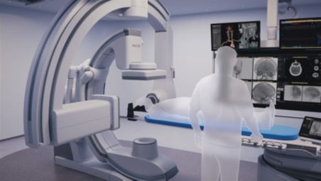
Secure and fast parking
Secure and fast parking of the lateral gantry allows for a seamless switch between 2D and 3D image acquisition. - Easily capture patient-oriented images from every angulation
-
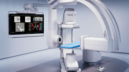
Easily capture patient-oriented images from every angulation
Image Beam Rotation guarantees patient-oriented images in every angulation. The functionality enables easy radial access due to the fact that the image is always aligned with the anatomical structure, even when positioned diagonally. No need to pivot the table or re-position the patient. - Full-body coverage
-
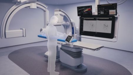
Full-body coverage
The system geometry combines speed and sophistication, enabling full-body coverage and giving you the most flexible Philips Azurion biplane to date. - Optimal access to the patient
-
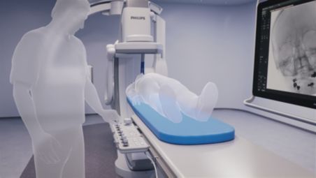
Optimal access to the patient
Positioning of the frontal arc at 135 degrees enables optimal head-end access to the patient and accommodates ideal working positions, for example, to free up the head-end for the anesthesiologist and optimal positioning of other medical equipment (e.g., ultrasound). - SmartCT Soft Tissue Helical*
-
SmartCT Soft Tissue Helical*
Improves neuro CBCT images to identify soft tissue changes in the Angio suite. An advanced protocol with dual-axis acquisition trajectory and improved reconstruction software results in improved image appearance, compared to conventional CBCT acquisition techniques. - Fuse 3D datasets for guidance during the procedure with SmartCT Dual Viewer*
-
Fuse 3D datasets for guidance during the procedure with SmartCT Dual Viewer*
SmartCT Dual Viewer offers 3D fusion imaging of 3D-RA and CBCT datasets during the procedure to support assessment and diagnosis. Load any two volumes from Azurion and compare at tableside without breaking sterility or switching applications.
Secure and fast parking
Secure and fast parking
Secure and fast parking
Easily capture patient-oriented images from every angulation
Easily capture patient-oriented images from every angulation
Easily capture patient-oriented images from every angulation
Full-body coverage
Full-body coverage
Full-body coverage
Optimal access to the patient
Optimal access to the patient
Optimal access to the patient
SmartCT Soft Tissue Helical*

SmartCT Soft Tissue Helical*
SmartCT Soft Tissue Helical*
Fuse 3D datasets for guidance during the procedure with SmartCT Dual Viewer*

Fuse 3D datasets for guidance during the procedure with SmartCT Dual Viewer*
Fuse 3D datasets for guidance during the procedure with SmartCT Dual Viewer*
- Secure and fast parking
- Easily capture patient-oriented images from every angulation
- Full-body coverage
- Optimal access to the patient
- Secure and fast parking
-
Secure and fast parking
Secure and fast parking of the lateral gantry allows for a seamless switch between 2D and 3D image acquisition. - Easily capture patient-oriented images from every angulation
-
Easily capture patient-oriented images from every angulation
Image Beam Rotation guarantees patient-oriented images in every angulation. The functionality enables easy radial access due to the fact that the image is always aligned with the anatomical structure, even when positioned diagonally. No need to pivot the table or re-position the patient. - Full-body coverage
-
Full-body coverage
The system geometry combines speed and sophistication, enabling full-body coverage and giving you the most flexible Philips Azurion biplane to date. - Optimal access to the patient
-
Optimal access to the patient
Positioning of the frontal arc at 135 degrees enables optimal head-end access to the patient and accommodates ideal working positions, for example, to free up the head-end for the anesthesiologist and optimal positioning of other medical equipment (e.g., ultrasound). - SmartCT Soft Tissue Helical*
-
SmartCT Soft Tissue Helical*
Improves neuro CBCT images to identify soft tissue changes in the Angio suite. An advanced protocol with dual-axis acquisition trajectory and improved reconstruction software results in improved image appearance, compared to conventional CBCT acquisition techniques. - Fuse 3D datasets for guidance during the procedure with SmartCT Dual Viewer*
-
Fuse 3D datasets for guidance during the procedure with SmartCT Dual Viewer*
SmartCT Dual Viewer offers 3D fusion imaging of 3D-RA and CBCT datasets during the procedure to support assessment and diagnosis. Load any two volumes from Azurion and compare at tableside without breaking sterility or switching applications.
Secure and fast parking
Secure and fast parking
Secure and fast parking
Easily capture patient-oriented images from every angulation
Easily capture patient-oriented images from every angulation
Easily capture patient-oriented images from every angulation
Full-body coverage
Full-body coverage
Full-body coverage
Optimal access to the patient
Optimal access to the patient
Optimal access to the patient
SmartCT Soft Tissue Helical*
SmartCT Soft Tissue Helical*
SmartCT Soft Tissue Helical*
Fuse 3D datasets for guidance during the procedure with SmartCT Dual Viewer*
Fuse 3D datasets for guidance during the procedure with SmartCT Dual Viewer*
Fuse 3D datasets for guidance during the procedure with SmartCT Dual Viewer*
Related products
Alternative products
-
SmartCT
- Simplifies 3D acquisition to empower clinical users to easily perform 3D imaging
- Enriches our outstanding 3D interventional tools with clear guidance
- 3D images are automatically displayed within seconds on the touch screen module
- Easily control and interact with advanced 3D visualization and measurement tools
View product
-
FlexSpot
- All relevant applications on one workspot for an efficient, clutter-free control room
- Includes one or two widescreen monitors, a mouse and a keyboard for full control
- Team members can perform all relevant tasks at a flexible workspot, without interrupting each other
- Create an unlimited number of screen layouts for every procedure and/or clinical user
View product
-
SmartCT Soft Tissue Helical
- Fast 10 secs helical trajectory acquisition to reduce motion artifacts
- Reconstruction software with cone beam, ring artifacts, bone beam, table scatter & vibration filters
- Automatic motion compensation functionality to salvage a CBCT with motion artifacts
- Metal artifact reduction algorithm to remove metal artifacts from the CBCT volume
- Workflow guidance with isocentering tool to aid 3D scan preparation
View product
-
Touch screen module pro
- Touch screen at table side for adjustments of imaging parameters without leaving the sterile field
- Collimate with a fingertip and zoom X-ray and roadmap images to support easy navigation
- View and control applications, as well as interventional tools and analysis applications
- A large mouse pointer can be made visible on your live image in the exam and control room
View product


-
SmartCT
Philips Image Guided Therapy clinical application software SmartCT enriches our outstanding 3D interventional tools with clear guidance, designed to remove barriers to acquiring 3D images in the interventional lab. Once acquired, 3D images are automatically displayed within seconds on the touchscreen module in the corresponding rendering mode. On the same touchscreen, the user can easily control and interact with advanced 3D visualizations and measurement tools. SmartCT also brings advanced measurements and visualization to your fingertips for high image quality, supporting your diagnosis[1-3] and better patient treatment outcomes[4-6].
View product
-
FlexSpot
As more applications come into your lab, it's more important than ever to work as efficiently as possible. FlexSpot gives you seamless access to all applications at one compact, customizable workplace to significantly reduce clutter and simplify workflow. Team members can perform different tasks separately, without interrupting each other, to reduce gaps between cases.
View product
-
SmartCT Soft Tissue Helical
SmartCT Soft Tissue Helical creates CBCT images to help spot soft tissue changes in the Angio suite. The new protocol with dual-axis acquisition trajectory and improved reconstruction software results in images with improved image appearance compared to conventional cone beam acquisition techniques. SmartCT Soft Tissue Helical is our improved CBCT protocol for neurovascular care with a fast 8 secs trajectory, metal artifact and motion compensation algorithms to further improve image quality.
View product
See all related products
- *SmartCT R3, and Azurion R3 are subject to regulatory clearance and may not be available in all markets. Contact your sales representative for more details.