FieldStrength MRI magazine
Research - May 2015
New insights for neuroscience
Researchers at Université de Sherbrooke (Sherbrooke, Quebec, Canada) are using techniques like denoising, advanced tractography, and simultaneous EEG-fMRI to better understand basic brain function in health and disease.
“Using a technique to denoise DWI allows push to connectome-project-like resolution in 13 minutes”
Maxime Descoteaux, PhD
Maxime Descoteaux, PhD, professor of computer science, director of the Imaging and Visualization Platform (PAVI) and director of the Sherbrooke Connectivity Imaging Laboratory (SCIL), is a leader in medical image analysis and image processing and an expert in diffusion magnetic resonance imaging acquisition, processing and visualization to infer white matter connectivity of the brain.
Kevin Whittingstall, PhD
Kevin Whittingstall, PhD, is an assistant professor in the department of Radiology at the Université de Sherbrooke in Québec, Canada and a Canada Research Chair in Neurovascular Coupling. His main research interests are in the development of non-invasive tools for measuring and interpreting brain function and structure in humans and animal models. In particular, his lab focuses on the balance between neural activity and cerebral blood supply (neurovascular coupling) and how disruptions in this balance are related to diseases of the brain (e.g. brain tumors).
Local and global challenge
Maxime Descoteaux, PhD, director of the Sherbrooke Connectivity Imaging Laboratory (SCIL), focuses on neuro connectivity, including algorithm development, modeling, and the processing pipeline. He notes two major challenges for neural imaging: understanding white matter microstructure and understanding the large scale connections in the brain. “At the local level, the challenge is to understand what the MR signal in a single voxel represents, especially in white matter. We have thousands of axons, blood vessels, and other types of cells such as glial cells, and all of this is averaged out in one MR signal, so there is a lot of room for local modeling to extract meaningful features from these signals,” Dr. Descoteaux notes. “The ultimate goal is to find new biomarkers for certain diseases such as neurodegenerative diseases, autism and psychiatric diseases.” “At a more global level, one of the biggest challenges is mapping the human connectome. As we map the connections of the brain, it brings so much data that we need new algorithms to analyze that data.”
Fast, high resolution DWI
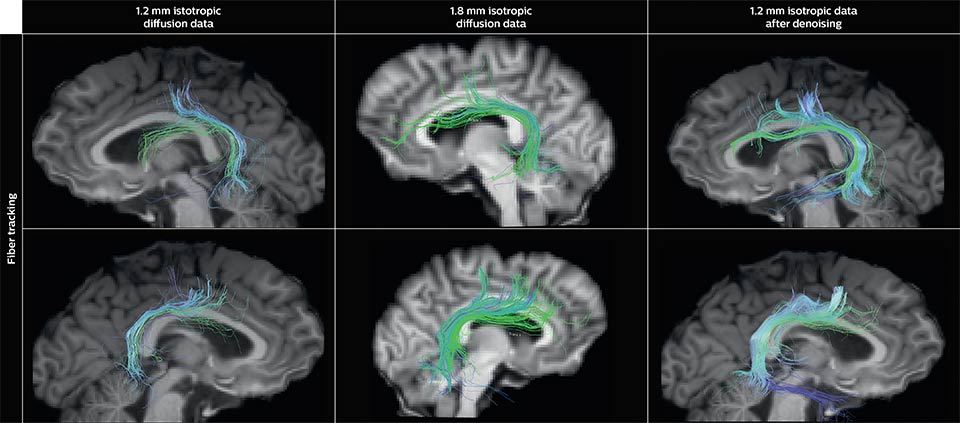
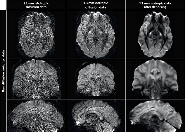
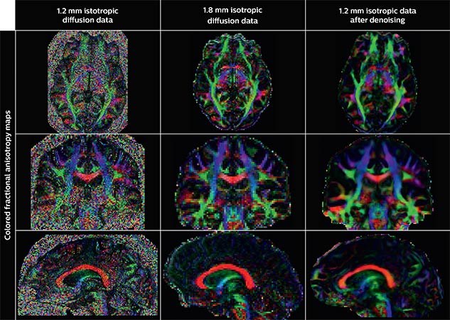
One of Dr. Descoteaux’s current projects involves using mathematical modeling and smart algorithms to optimize diffusion weighted images (DWI). He explains that in DWI, as in all MR techniques, SNR decreases and acquisition time increases as the voxel size is reduced, forcing DWI acquisitions at a spatial resolution that can’t provide the desired high specificity of reconstructed tracts and diffusion features. Dr. Descoteaux and his colleagues have published a scientific paper at the ISMRM (2015) that concludes that applying his denoising techniques can produce acquisition of high resolution DWIs comparable to those acquired in the Human Connectome Project. “The difference is that our dataset was acquired in 13 minutes on a clinical 3.0T scanner without expensive, specialized hardware, as opposed to about an hour and a half on the Connectome Project systems,” he points out.
“The ultimate goal is to find new biomarkers for certain diseases such as neurodegenerative diseases, autism and psychiatric diseases”
Denoising to improve quality
Using a non-local spatial and angular block matching technique to denoise raw diffusion weighted images. allows to push acquisition to lower spatial resolution and read human-connectome-project-like resolution from standard Philips Ingenia 3.0T MRI scanner. The data were acquired with spatial resolution of 1.2 x 1.2 x 1.2 mm in 13 minutes for 40 full brain DWI with b 1000 and one with b 0
“More or bigger veins will inherently produce bigger BOLD responses. So you have to be careful when comparing two activation sites in a subject or among subjects”
Correcting for vascular density
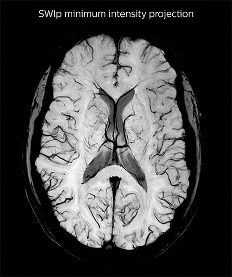
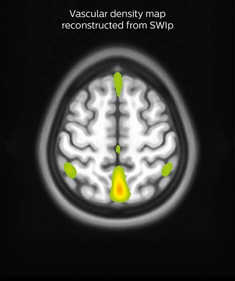
A SWIp image (left) is used to visualize veins in cortical and sub-cortical areas. Using in-house reconstruction techniques, a vascular density map is obtained in individual subjects and averaged over a population (right). Areas in red/green represent areas with dense venous vascularization. The lab uses such images to correct fMRI (BOLD) activation maps in order to minimize false positives.
Simultaneous EEG fMRI
Dr. Whittingstall is also interested in how EEG correlates with fMRI. “We’re investigating if changes in a certain EEG frequency band better explain the changes that we’re seeing in the BOLD response, or if it is some combination of multiple frequency bands from different brain areas,” he says. Neuroscience tools aid research Dr. Whittingstall uses iViewBold to map functional areas and connectivity. “iViewBold enables us to see immediately if a subject is properly doing a task, or if the subject is a worthy BOLD responder,” he explains. “The prospective motion correction is also very helpful. Usually, online fMRI analysis packages don’t always take online registration into account, but the Philips system does online correcting, which helps make fMRI maps more reliable.” In addition, together with Philips, the team is evaluating a new tool to display – in real time – the motion of the subject during the fMRI acquisition. The team uses the 32-channel dS Head coil for its studies. “With a very short TR of just below two seconds, we’re getting beautiful signal-to-noise ratio in the visual cortex using fairly subtle visual stimuli,” Dr. Whittingstall notes.
“There are many DICOM to NIfTI converters, but it is convenient to have it done at the console”
Both Dr. Whittingstall and Dr. Descoteaux appreciate that the neuroscience package makes it easy to convert DICOM images to a NIfTI format. “There are many DICOM to NIfTI converters,” Dr. Whittingstall says, “but it is convenient to have it done at the console.” Through a research agreement with Philips, the researchers also have access to the Paradise pulse programming environment and sequence simulator. “It’s like a virtual scanner,” Dr. Descoteaux explains. “I can prepare my protocols from home and then I’m set up and ready as soon as I get to the scanner.” Reproducibility impresses Drs. Descoteaux and Whittingstall chose a digital Philips Ingenia MR system after downloading and analyzing the source data from scans on different systems. “It’s great to publish your findings, but ultimately, you want to make sure that they’re reproducible, and access to the source data is the only way we can see exactly what is happening,” Dr. Whittingstall says. Dr. Descoteaux adds that they preferred Philips performance in the categories reviewed, including SNR, fMRI stability, and the angular resolution for diffusion data.
“Ultimately, we want to make sure that data are reproducible, and access to the source data is the only way we can see exactly what is happening”
References
1. St-Jean S, Gilbert F, Descoteaux M Connectome -like quality diffusion MRI in 13 minutes - Improving diffusion MRI spatial resolution with denoising ISMRM 2015
2. Vigneau-Roy N, Bernier M, Descoteaux M, Whittingstall K Regional variations in vascular density correlate with resting-state and task-evoked blood oxygen level- dependent signal amplitude Human Brain Mapping 2014, 35 (5), 1906-192
More from FieldStrength
